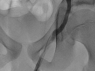
Bag of worms
Nice fluoroscopic image of contrast stasis in a varicocele and left spermatic vein after descending flebography in a 12 year old boy. This actually isn’t […]

Nice fluoroscopic image of contrast stasis in a varicocele and left spermatic vein after descending flebography in a 12 year old boy. This actually isn’t […]
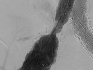
Pre and post image of a trombotic stenosis in a Cook! Zenith Alpha endoprosthesis. It was relined with a 16×60 mm Wallstent.
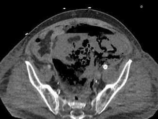
A 74 year old male patient was referred to our hospital with a ruptured abdominal aortic aneurysm (figures 1a-d). In an emergency setting using only […]
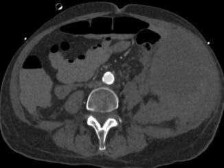
On the left two slices showing a huge hematoma in the left flank. On the right the same two slices 4 months later. One might […]
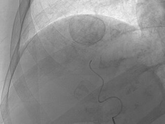
Certainly not a true ‘evil eye’ but still another nice post chemoembolization image outlining the HCC in liver segment 7 through captivation of contrast on […]
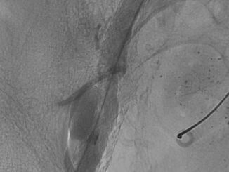
Three cases of stent related aneurysms, just as a friendly reminder that stents are not always harmless. The last case has an impressive solution. Case […]
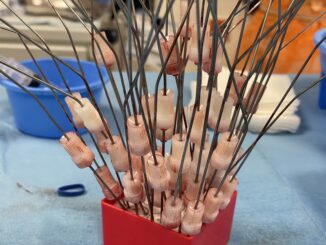
We call this a ‘hedgehog’. It refers to the look of the temporary sharps holder after a really intensive coil embolization.
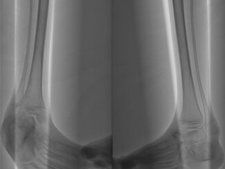
Angiography of the lower legs in a patient with a rare syndrome. So rare that we will not mention it here, in fear of the […]
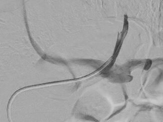
Images of biochemically confirmed left adrenal veins. Use your HI (Human Intelligence) to create a reference for future procedures. On the AP view the confluence […]
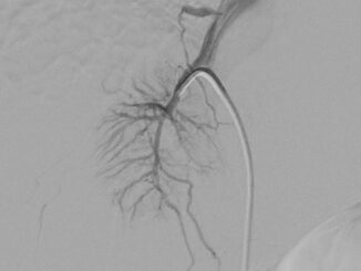
Images of biochemically confirmed rightsided adrenal veins. Use your HI (Human Intelligence) to create a reference for future procedures. Typical for the right adrenal vein […]
Copyright © 2025 | WordPress Theme by MH Themes