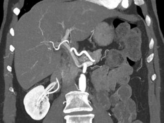
The Liver Divided II
This is a coronal MIP reformatted longitudinally along the portal vein. Interestingly there is contrast rich blood in the left portal vein but contrast poor […]

This is a coronal MIP reformatted longitudinally along the portal vein. Interestingly there is contrast rich blood in the left portal vein but contrast poor […]
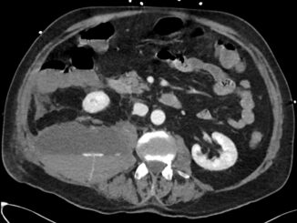
Nice image of a (right sided) psoas hematoma with active contrast extravasation. The appearance of a fluid level is almost pathognomic for anticoagulation therapy, precipitating […]
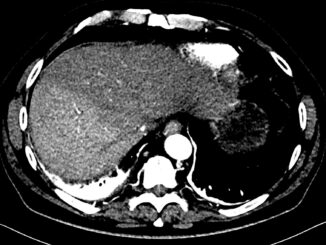
Three CT images of the liver at the same level: unenhanced, arterial phase and venous phase. In the arterial phase the liver is divided along […]
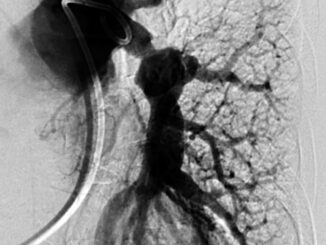
In the workup for chronic thromboembolic pulmonary hypertension (CTEPH) this 73 year old female patient was referred to the interventional radiology department for pulmonary angiography. […]
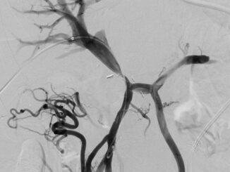
Nice phlebograph of the portal system showing portal vein stenosis (near the operation clips). The angiographic catheter was placed transhepatically in the superior mesenteric vein. […]
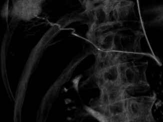
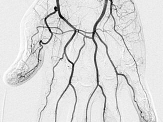
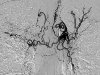
Angiography of a left bronchial artery in a 20 year old patient with cystic fibrosis. There are numerous pathological bronchial arteries visible. There are also […]
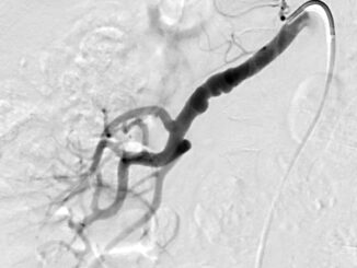
Typical image of incidentally found fibromuscular dysplasia (FMD) of the renal artery with short stenoses interspersed with short dilated segments (this patient was screened for […]
Copyright © 2025 | WordPress Theme by MH Themes